What goes wrong anatomically
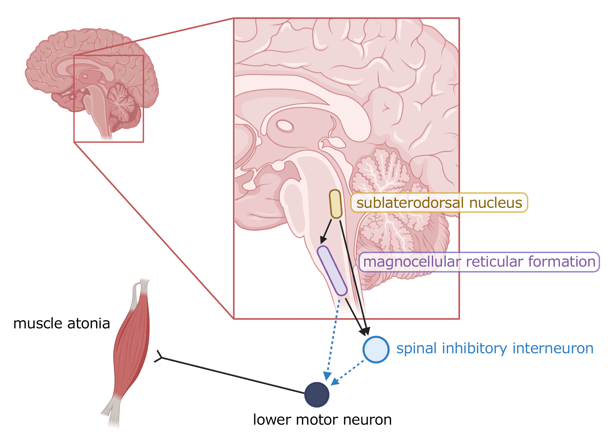
During healthy REM sleep, a small cluster of neurons located in the brainstem actively paralyzes the body's muscles. This state of transient paralysis requires continuous inhibitory signal sent downward to the spinal cord to prevent movement during dreaming. In RBD, this system breaks down. Motor commands generated by the dreaming brain can reach the muscles unopposed, and the body is able to move, or act out dreams.
Why the circuit fails
In most adults who develop RBD without an obvious trigger, the cause is the gradual accumulation of misfolded alpha-synuclein protein in the brainstem (pons, medulla). This is the same protein pathology that underlies Parkinson's disease and Lewy body dementia — and the circuits that control REM atonia are among the first structures it reaches, often years or decades before other symptoms appear. Other causes exist, particularly in younger adults: certain antidepressants (SSRIs and SNRIs) can pharmacologically suppress REM atonia; and rare autoimmune conditions affecting the brain can damage the same circuitry through inflammation.

How the diagnosis is made
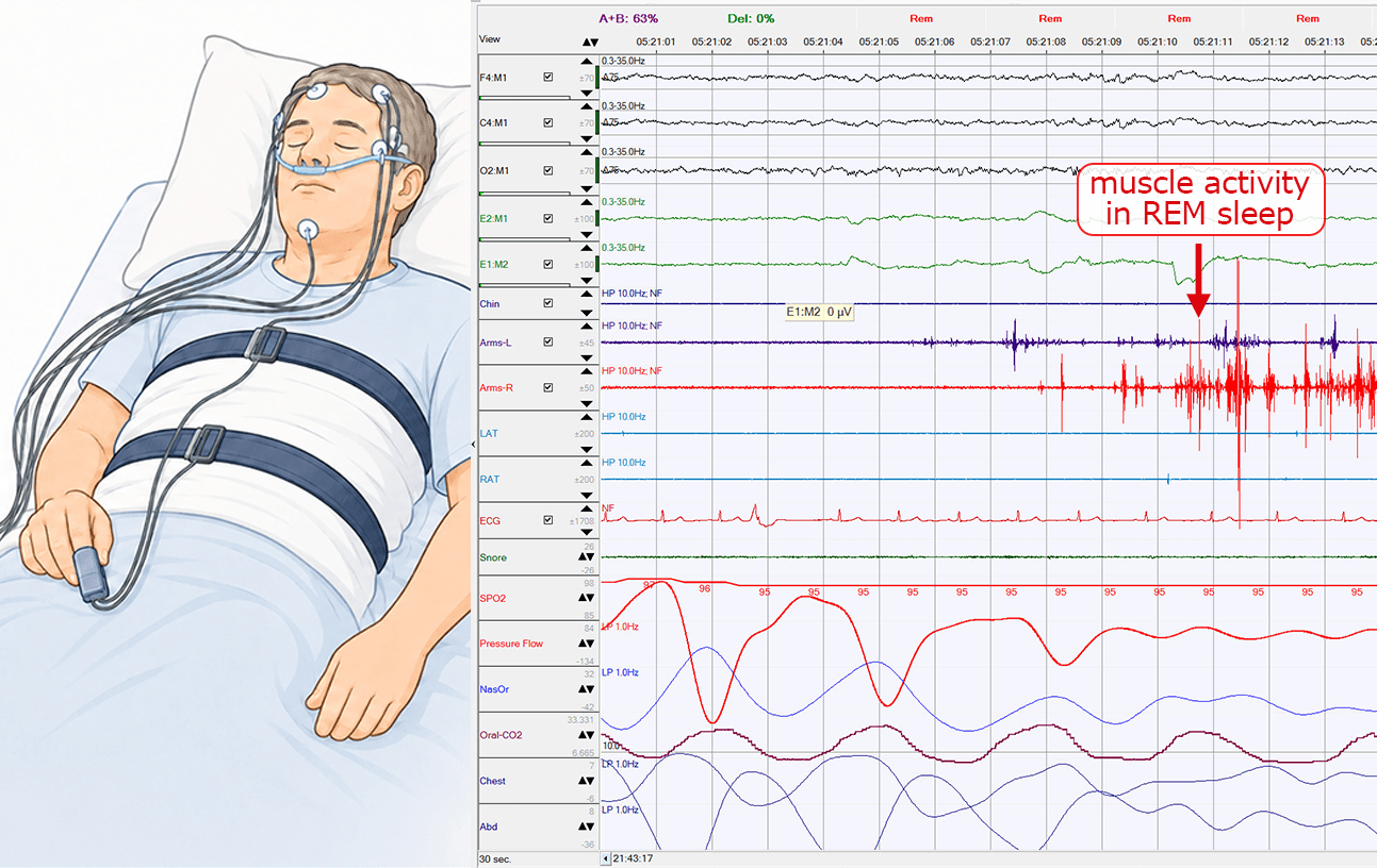
Questionnaires can flag people who may have RBD, but they lack specificity because other sleep conditions can present similarly, including sleep apnea, leg movements during sleep, and other parasomnias that occur in the other stage called non-REM sleep. The gold standard is polysomnography, a single overnight recording in a sleep laboratory that measures brain activity, muscle tone, eye movements, and video simultaneously. What the clinician is looking for is “REM sleep without atonia” — the loss of normal muscle paralysis — ideally accompanied by observed behavior. That very specific combination confirms the diagnosis. But polysomnography is expensive, resource-intensive, not widely accessible, and the test can miss the diagnosis based on a single night of recording. This program aims to expand diagnosis by leveraging the scalability and accuracy of wearable-based algorithms to identify people at high risk of RBD.